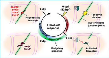
Despite their importance in tissue maintenance and repair, fibroblast diversity and plasticity remain poorly understood. Using single-cell RNA sequencing, University of Calgary researchers uncover distinct sclerotome-derived fibroblast populations in zebrafish, including progenitor-like perivascular/interstitial fibroblasts, and specialized fibroblasts such as tenocytes. To determine fibroblast plasticity in vivo, the researchers develop a laser-induced tendon ablation and regeneration model. Lineage tracing reveals that laser-ablated tenocytes are quickly regenerated by preexisting fibroblasts. By combining single-cell clonal analysis and live imaging, the researchers demonstrate that perivascular/interstitial fibroblasts actively migrate to the injury site, where they proliferate and give rise to new tenocytes. By contrast, perivascular fibroblast-derived pericytes or specialized fibroblasts, including tenocytes, exhibit no regenerative plasticity. Active Hedgehog (Hh) signaling is required for the proliferation of activated fibroblasts to ensure efficient tenocyte regeneration. Together, this work highlights the functional diversity of fibroblasts and establishes perivascular/interstitial fibroblasts as tenocyte progenitors that promote tendon regeneration in a Hh signaling-dependent manner.
scRNA-seq reveals heterogeneity and differential plasticity in sclerotome-derived fibroblasts
(A) Sample preparation pipeline for scRNA-seq of mCherry+ cells from 52 hpf nkx3-1NTR-mCherry zebrafish trunks. (B) UMAP plot of the post-filtering scRNA-seq dataset. Dotted line indicates sclerotome-derived fibroblast clusters. (C) Proportion of ECM-related (matrisomal) transcripts detected per cell, grouped by cluster. Matrisome gene expression is enriched in sclerotome-derived fibroblast clusters (dotted line). (D) UMAP representation of subsetted fibroblast clusters from (B), with distinct fibroblast subtypes labeled. (E) Feature plots showing expression of a pan-fibroblast marker, pdgfra, and one marker for each cluster identified in (D). (F) Schematic depicting distribution of fibroblast subtypes from (D) in the zebrafish trunk, as determined in figs. S4 to S6. (G) RNA velocity analysis of fibroblast clusters. Stream and arrowheads indicate direction of differentiation trajectory. CVP, caudal vein plexus; DLAV, dorsal longitudinal anastomotic vessel; Fb, fibroblast; ISV, intersegmental vessel; MTJ, myotendinous junction; Scl, sclerotome.
Rajan AM, Rosin NL, Labit E, Biernaskie J, Liao S, Huang P. (2023) Single-cell analysis reveals distinct fibroblast plasticity during tenocyte regeneration in zebrafish. Sci Adv 9(46):eadi5771. [article]


