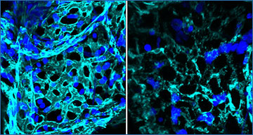
Pulmonary fibrosis develops as a consequence of failed regeneration after injury. Analyzing mechanisms of regeneration and fibrogenesis directly in human tissue has been hampered by the lack of organotypic models and analytical techniques.
Researchers at Helmholtz Munich have coupled ex vivo cytokine and drug perturbations of human precision-cut lung slices (hPCLS) with single-cell RNA sequencing and induced a multilineage circuit of fibrogenic cell states in hPCLS. The researchers showed that these cell states were highly similar to the in vivo cell circuit in a multicohort lung cell atlas from patients with pulmonary fibrosis. Using micro-CT-staged patient tissues, they characterized the appearance and interaction of myofibroblasts, an ectopic endothelial cell state, and basaloid epithelial cells in the thickened alveolar septum of early-stage lung fibrosis. Induction of these states in the hPCLS model provided evidence that the basaloid cell state was derived from alveolar type 2 cells, whereas the ectopic endothelial cell state emerged from capillary cell plasticity. Cell-cell communication routes in patients were largely conserved in hPCLS, and antifibrotic drug treatments showed highly cell type-specific effects. This work provides an experimental framework for perturbational single-cell genomics directly in human lung tissue that enables analysis of tissue homeostasis, regeneration, and pathology. The researchers further demonstrate that hPCLS offer an avenue for scalable, high-resolution drug testing to accelerate antifibrotic drug development and translation.
An ex vivo model of human lung fibrogenesis recapitulates early-stage events in PF patients
(a) Benchmarking strategy: Human precision-cut lung slices (hPCLS) were generated from uninvolved peritumoral tissue and treated under the indicated conditions before single cell RNA-seq analysis (FC = Fibrotic Cocktail: TGFb1, PDGFb, TNFa, and LPA, CC = Control Cocktail: diluents of the components) at day six of treatment. This data was benchmarked against in vivo transcriptome data from PF patients as indicated. (b, c) UMAP embedding of 63,581 single cells from the hPCLS model, color coded by cell type (b) and treatment (c), respectively. (d, e) UMAP embedding of 481,788 single cells from the integrated multi-cohort PF cell atlas, color coded by cell type (d) and disease status (e), respectively. (f) Ex vivo marker genes signatures of meta-cell types in hPCLS. The heatmap shows the average scaled expression of markers in each meta-cell type. (g) matchSCore comparison of ex vivo marker genes against in vivo marker genes. (h) Query-to-reference mapping with scArches overview: ex vivo hPCLS and in vivo PF scRNA-seq data were mapped to the Human Lung Cell Atlas (HLCA) reference for benchmarking and systematic side-by-side comparisons. (i) Similarity of CC and FC treated cells from hPCLS compared to cells from Control and Pulmonary Fibrosis in the PF-extended HLCA (in vivo reference) as assessed by scArches mapping. Stacked bar plots show the percentage of cells mapping to either cells from healthy controls or PF patients with regards to CC or FC treatment for each cell type. Fisher’s exact test. (j) IPF stage-specific signatures derived from publicly available bulkRNA-seq data25. The venn diagram illustrates the intersection of differentially upregulated genes in IPF stages 1, 2 and 3 (versus control tissue). Selected genes from these signatures and enriched GO terms are highlighted. (k) Comparison of hPCLS and IPF stage-specific signatures. The bar plot shows the overlap of differently upregulated genes (in %) between the in vivo stage-specific IPF signatures and the ex vivo hPCLS signature (FC vs. CC).
Lang NJ, Gote-Schniering J, Porras-Gonzalez D, Yang L, De Sadeleer LJ, Jentzsch RC, Shitov VA, Zhou S, Ansari M, Agami A, Mayr CH, Hooshiar Kashani B, Chen Y, Heumos L, Pestoni JC, Molnar ES, Geeraerts E, Anquetil V, Saniere L, Wögrath M, Gerckens M, Lehmann M, Yildirim AÖ, Hatz R, Kneidinger N, Behr J, Wuyts WA, Stoleriu MG, Luecken MD, Theis FJ, Burgstaller G, Schiller HB. (2023) Ex vivo tissue perturbations coupled to single-cell RNA-seq reveal multilineage cell circuit dynamics in human lung fibrogenesis. Sci Transl Med 15(725):eadh0908. [abstract]


