

Amyotrophic lateral sclerosis (ALS) is a challenging and progressive neurodegenerative disease. It primarily affects motor neurons, the cells responsible for controlling our muscles, leading to a loss of motor function over time. Despite extensive research, scientists still grapple with understanding why certain neurons are more susceptible to damage in ALS. A recent study led by researchers at Harvard University sheds light on this mystery by delving into the molecular characteristics of these vulnerable neurons.
The Focus on ETNs
The study zeroes in on a specific group of neurons known as extratelencephalic neurons (ETNs), including Betz cells. These neurons play a critical role in motor function, and their degeneration is a hallmark of ALS. Researchers sought to understand what makes ETNs particularly prone to ALS-related damage.
Investigating the Neurons: RNA Sequencing
To uncover the unique traits of ETNs, scientists used a technique called RNA sequencing. This method allows them to analyze the genetic activity within individual cells. In this study, they examined over 79,000 single nuclei from the brains of both ALS patients and healthy individuals. This comprehensive analysis provided insights into the differences in gene expression between these groups.
Key Findings
- Higher Expression of ALS Risk Genes:
- The study found that ETNs, whether from ALS patients or healthy individuals, had a higher expression of genes associated with ALS risk. This suggests that these neurons naturally have a predisposition towards ALS due to their genetic makeup.
- Stress Responses in ALS Patients:
- In the ETNs of ALS patients, there was a notable increase in the activity of genes involved in managing protein homeostasis and stress responses. These changes indicate that ETNs in ALS patients are under significant stress, potentially contributing to their degeneration.
- Changes in Support Cells:
- The study also looked at oligodendrocytes (cells that insulate neurons) and microglia (immune cells in the brain). In ALS patients, oligodendrocytes showed a decrease in genes related to myelination (the process of forming the protective sheath around neurons). Microglia, on the other hand, exhibited an increase in genes associated with an endolysosomal reactive state, indicating a heightened response to cellular stress and damage.
Cellular susceptibility to ALS–FTD in the human postmortem cortex
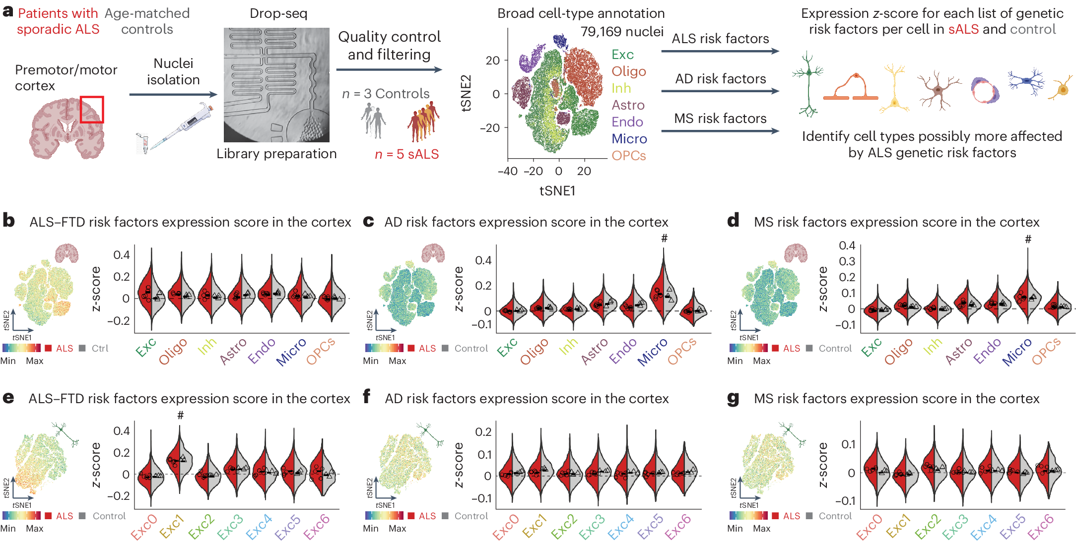
a, Diagram of the workflow for isolation of nuclei from cortices of patients with ALS and age-matched controls followed by snRNAseq and assessment of expression of gene modules associated with neurodegenerative diseases. Exc, excitatory neurons; Inh, inhibitory neurons; Oligo, oligodendrocytes; OPCs, oligodendrocyte progenitor cells; Micro, microglia; Astro, astrocytes; Endo, endothelial cells. b–d, tSNE projections and violin plots of z-scores for expression of genes associated with ALS–FTD (b), AD (c) and MS (d) in the different cell types identified (the bars denote median for each side of the violin and the symbols indicate the average score per individual). e–g, tSNE projections and violin plots of z-scores for expression of genes associated with the ALS–FTD (e), AD (f) and MS (g) in the different subtypes of excitatory neurons (the bars denote median for each side of the violin and the symbols indicate the average score per individual) (in a–g, n = 3 control individuals and n = 5 patients with sALS).
Implications of the Findings
These findings suggest that the vulnerability of ETNs in ALS is linked to their intrinsic molecular properties. The higher expression of ALS risk genes makes them more sensitive to the genetic and environmental factors that drive ALS. Additionally, the stress responses observed in ALS patients’ ETNs highlight how these neurons struggle under disease conditions, leading to their degeneration.
The changes in oligodendrocytes and microglia also point to a broader disruption in the brain’s cellular environment in ALS, further contributing to neuronal damage.
Conclusion
Understanding why ETNs are particularly susceptible to ALS offers critical insights into the disease’s progression. This knowledge not only helps explain the selective vulnerability of these neurons but also opens up potential avenues for targeted therapies aimed at protecting ETNs and managing stress responses. By continuing to unravel the complex molecular landscape of ALS, researchers move closer to developing effective treatments for this devastating disease.
Limone F, Mordes DA, Couto A, Joseph BJ, Mitchell JM, Therrien M, Ghosh SD, Meyer D, Zhang Y, Goldman M, Bortolin L, Cobos I, Stevens B, McCarroll SA, Kadiu I, Burberry A, Pietiläinen O, Eggan K. (2024) Single-nucleus sequencing reveals enriched expression of genetic risk factors in extratelencephalic neurons sensitive to degeneration in ALS. Nat Aging [Epub ahead of print]. [article]


